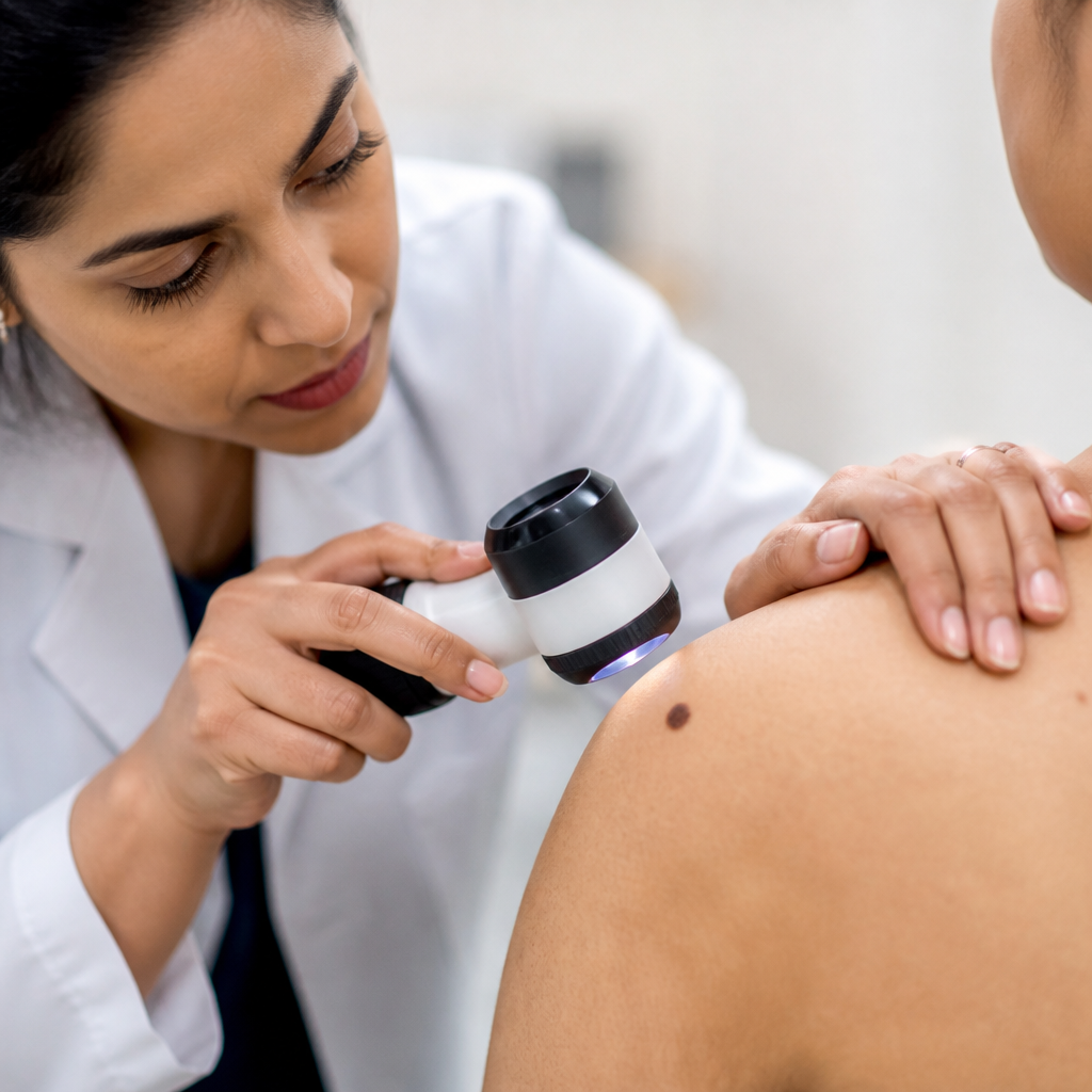
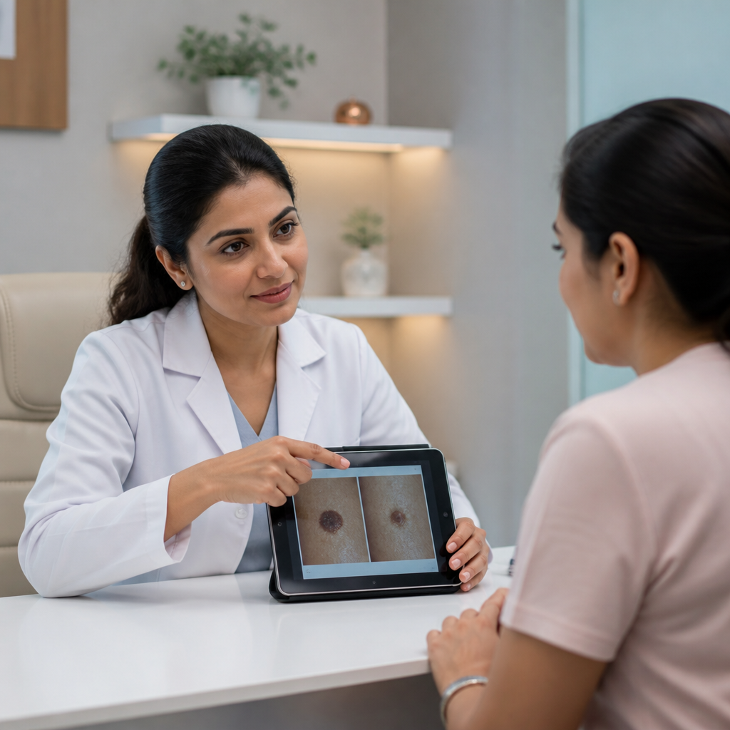
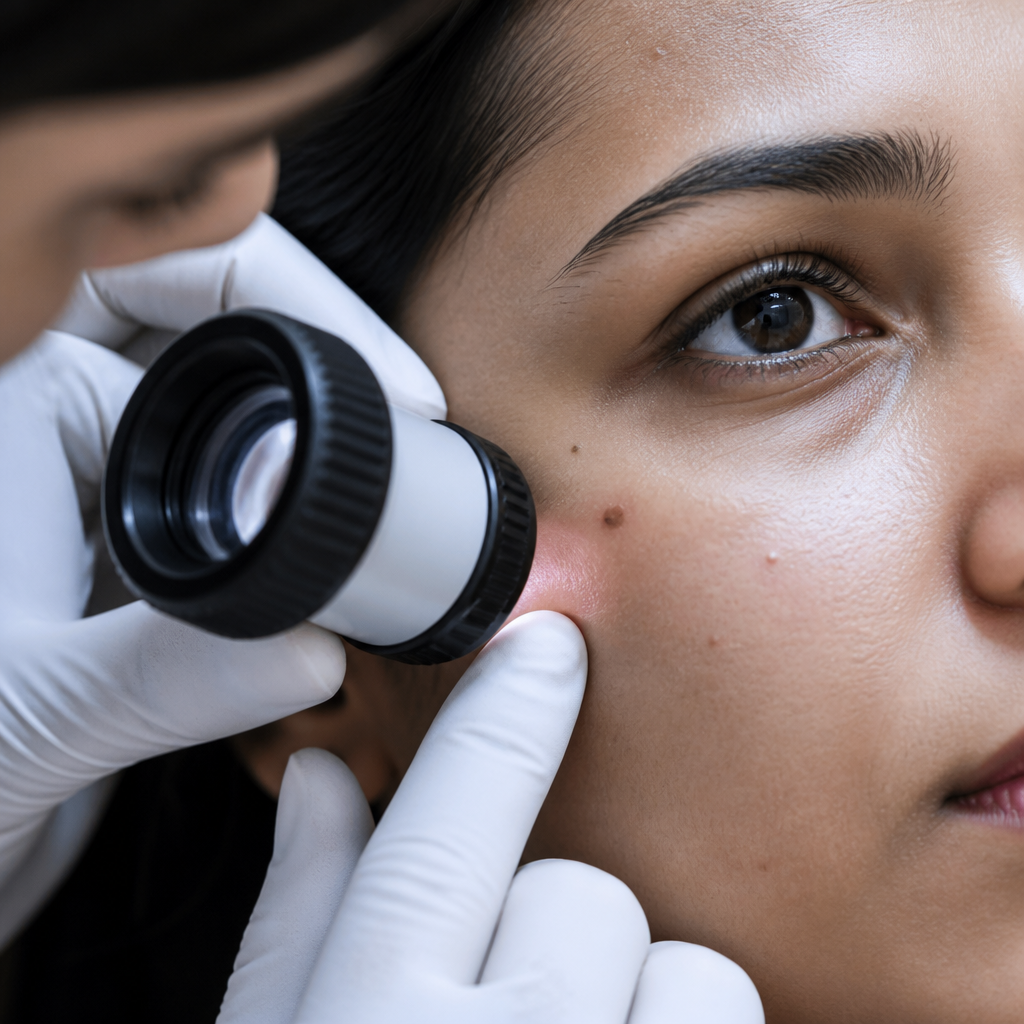
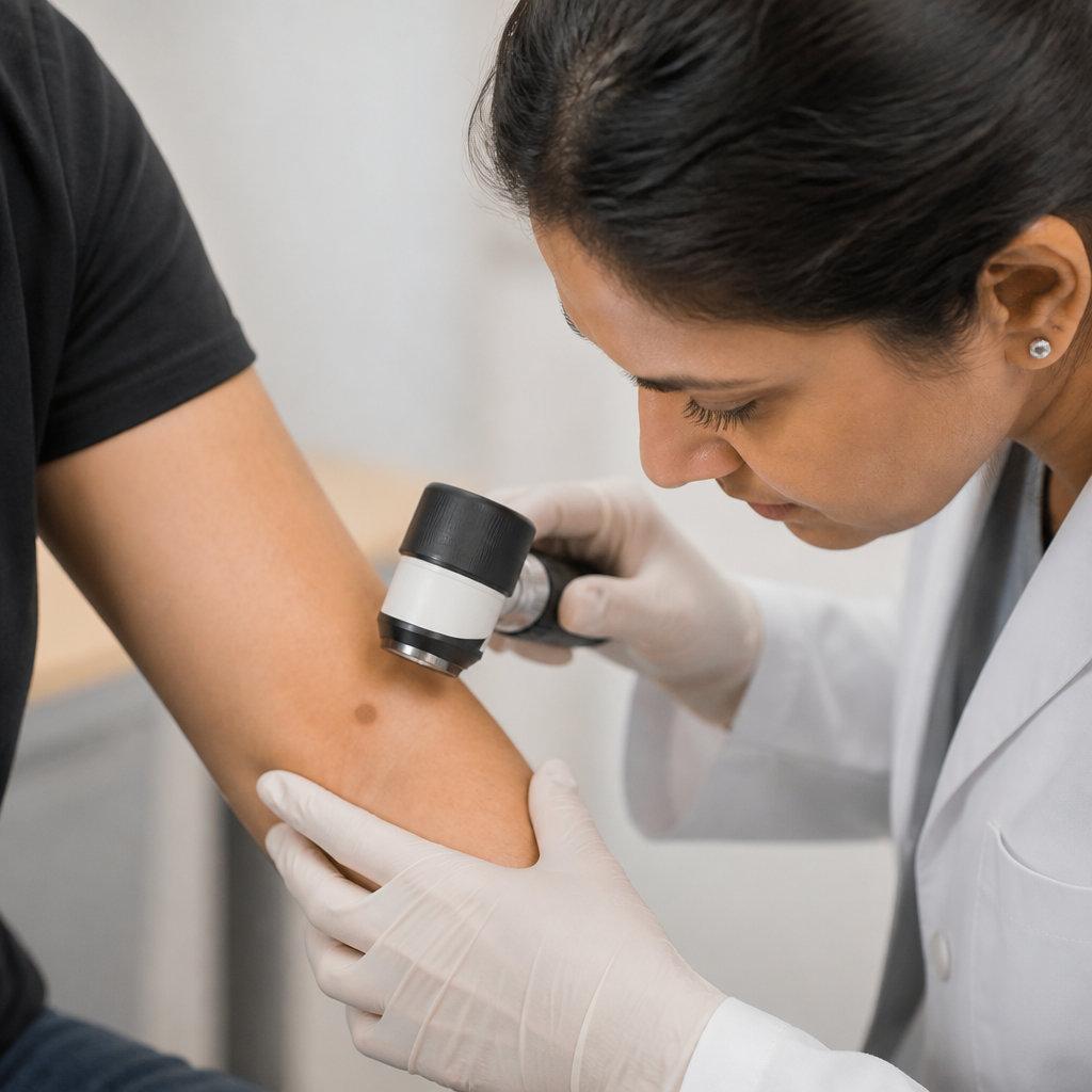
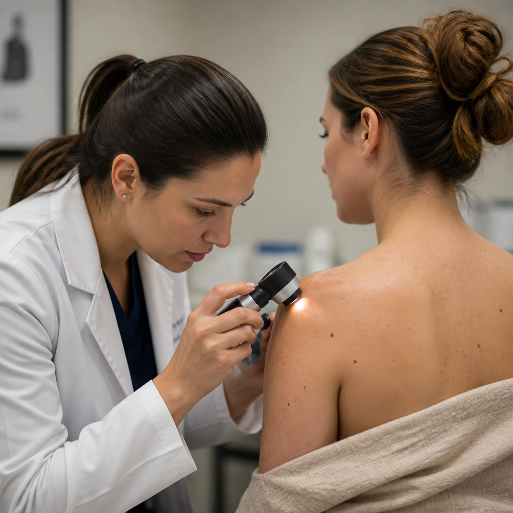
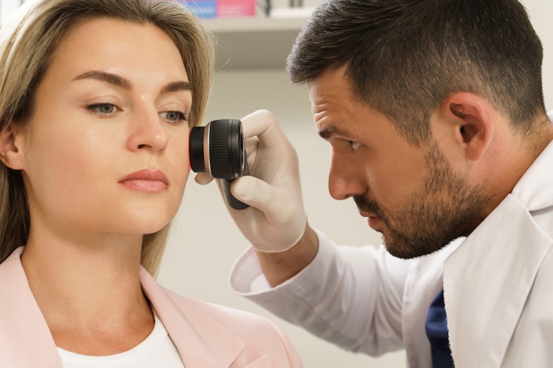
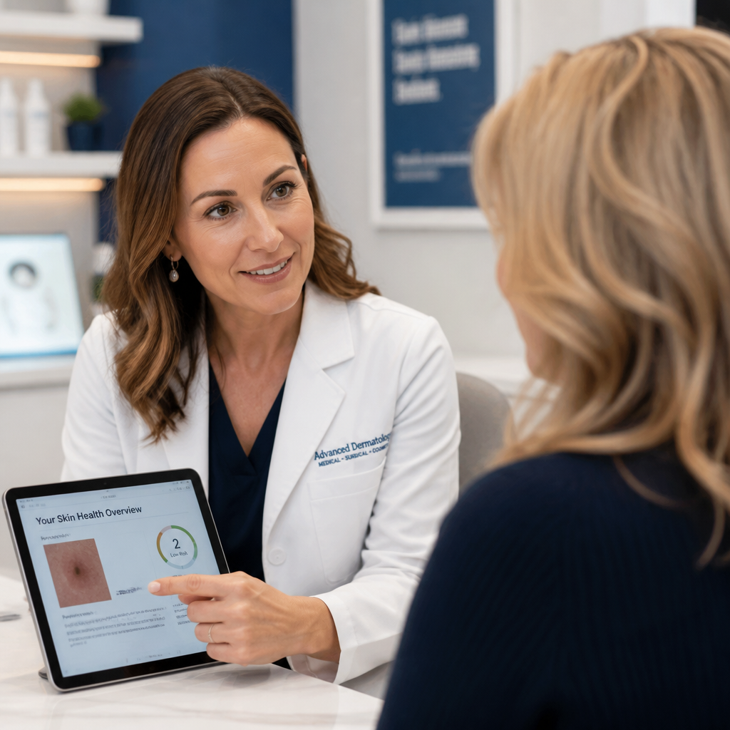
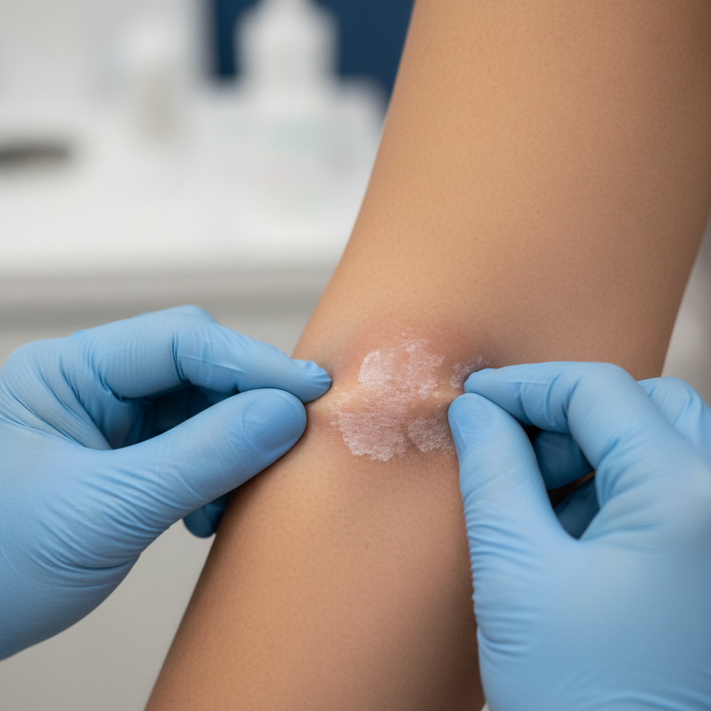
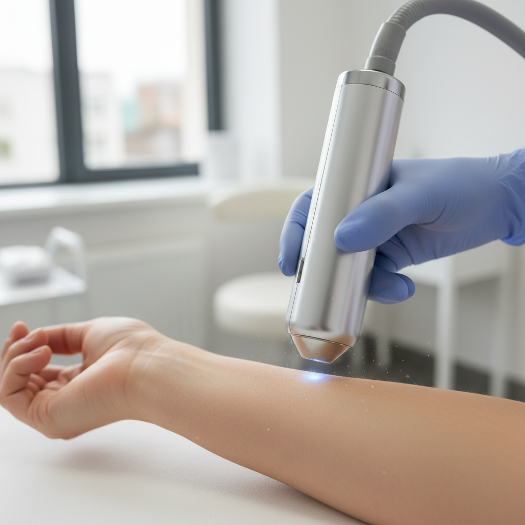
Early detection is the most powerful tool against skin cancer. Our senior dermatologists perform full-body mole mapping, dermoscopy, and targeted biopsies — identifying malignant lesions before they become dangerous.
From annual mole mapping for high-risk individuals to urgent evaluation of suspicious lesions — we provide the full spectrum of dermatological cancer screening.
Systematic, photographically documented examination of all moles under dermoscopy. Baseline images stored for year-on-year comparison — detecting change before it's clinically visible.
Any mole showing ABCDE changes examined under 10× polarised dermoscopy — achieving 90%+ diagnostic accuracy compared to 60–70% with clinical eye alone. Results documented same day.
The most dangerous skin cancer — curable when caught early. We perform dermoscopic evaluation and same-day excision biopsy of suspicious lesions, with histopathology at accredited labs.
The most common skin cancer — often a pearly nodule or non-healing sore on sun-exposed skin. We identify BCC under dermoscopy and perform excision or co-ordinate Mohs surgery referral.
Firm, keratotic lesions on scalp, ears, or lips. We biopsy, stage, and arrange appropriate surgical or radiation referral with complete co-ordination and follow-up throughout.
Patients with family history of melanoma, atypical moles, previous skin cancer, immunosuppression, or significant UV history receive personalised surveillance at 6–12 monthly intervals.
A complete skin cancer screening is thorough, systematic, and entirely painless — from history to dermoscopy to biopsy if needed, all in one appointment.
UV exposure history, family history of melanoma, number of moles, previous skin cancers, immunosuppressive medications, and skin type documented to guide screening intensity.
Head-to-toe examination under optimal lighting — including scalp, between toes, and under nails. Every site is examined methodically, with suspicious lesions flagged for dermoscopy.
Polarised light dermoscopy at 10× magnification applied to any lesion with concerning features — distinguishing benign from potentially malignant with >90% accuracy.
If dermoscopy shows suspicious features, same-day excision or punch biopsy performed under local anaesthetic. Specimen sent to accredited histopathology lab within 24 hours.
Histopathology results reviewed and explained at follow-up. Benign: added to mole map. Malignant: urgent surgical oncology referral facilitated with clear communication throughout.
Clinical eye alone misses up to 26% of melanomas. Dermoscopy with our experienced dermatologists achieves >90% accuracy — the standard of care for pigmented lesion evaluation.
Polarised light dermoscopy applied to every suspicious lesion — the evidence-based standard that clinical examination alone cannot match.
All moles photographed and stored at baseline — enabling precise year-on-year comparison to detect change before it becomes visible to the naked eye.
When dermoscopy raises concern, we perform excision or punch biopsy the same day — no delays, no waiting for a second appointment.
Malignant results are managed with urgency — we co-ordinate histopathology, staging, and surgical oncology referral with clear communication throughout.
| Feature | Dermoscopy Screening | Clinical Eye Alone | GP Referral | Self-Examination |
|---|---|---|---|---|
| Diagnostic accuracy | >90% | ~60–70% | Variable | <50% |
| Mole photography | ✓ Yes | No | No | No |
| Same-day biopsy | ✓ Yes | No | Weeks wait | No |
| High-risk surveillance | ✓ Personalised plan | Rarely | Standard intervals | Self-managed |
| Starting cost | ₹1,500 screening | N/A | GP consult fee | Free |
Each diagnostic tool is selected based on the clinical presentation and risk profile of each patient.
10× magnification polarised-light dermoscope distinguishes benign moles from early melanoma with >90% diagnostic accuracy.
Melanoma · BCC DetectionDigital photography of all moles at baseline, stored for annual comparison. Changes over time are the earliest signal of malignant transformation.
Annual SurveillanceSame-day biopsy under local anaesthesia. Full histopathological reporting with Breslow depth, Clark level, and margin status.
Same-Day BiopsyUV fluorescence identifies sub-clinical pigmented lesions and actinic damage — complementary to dermoscopy for comprehensive assessment.
UV MappingAt Supreme Elite Clinic, Kelambakkam OMR 603103
Early detection saves lives. Book a full-body dermoscopy screening — the single most important thing you can do if you have risk factors for skin cancer.
Book ScreeningCall +91 96774 73344 or fill the form below and we'll call you back within 2 hours.