Glaucoma Care

Glaucoma
Management

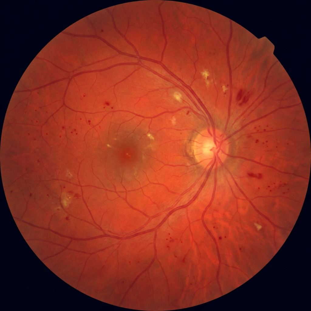
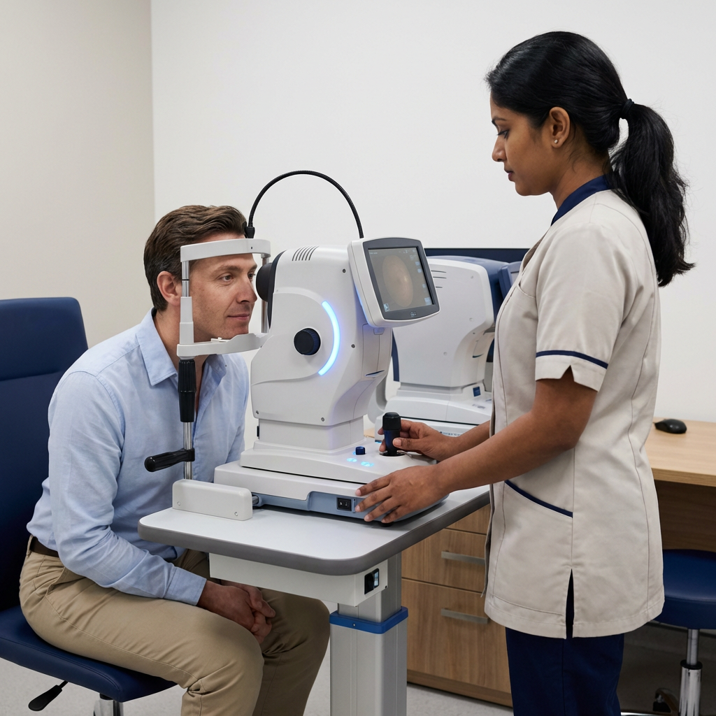
The "silent thief of sight" causes irreversible vision loss before you notice a single symptom. Early detection with OCT and visual field analysis — combined with targeted treatment — is the only way to preserve your vision for life.

0Symptoms in early stages
OCTNerve fibre imaging
SLTLaser — 5 min procedure
Glaucoma eye examination OCT
Annual screeningCritical for over-40s & diabetics
Types of Glaucoma

Understanding Your Diagnosis

Glaucoma is not a single disease — it's a group of conditions. The type determines the treatment approach and urgency.

Primary open angle glaucoma
Most Common

Primary Open-Angle Glaucoma (POAG)

Accounts for 70% of glaucoma. Drainage angle remains open but trabecular meshwork gradually loses efficiency — slowly raising IOP and damaging the optic nerve over years with no pain or visual symptoms until advanced.

Lifelong monitoringDrops / SLT / MIGS
Angle closure glaucoma acute
Acute Emergency

Angle-Closure Glaucoma

The iris physically blocks the drainage angle, causing sudden IOP spikes to 40–70 mmHg. Symptoms: severe eye pain, headache, nausea, haloes around lights. This is an ophthalmic emergency — requires same-day laser iridotomy.

Urgent treatmentYAG Iridotomy
Normal tension glaucoma
Atypical Type

Normal-Tension Glaucoma (NTG)

Optic nerve damage occurs despite IOP within the normal range (<21 mmHg). Vascular insufficiency to the optic nerve is implicated. OCT and visual field analysis are essential — IOP alone would miss this diagnosis entirely.

Low-pressure managementSpecialised drops
Ocular hypertension glaucoma risk
Pre-Glaucoma

Ocular Hypertension (OHT)

IOP above 21 mmHg with no optic nerve damage or visual field loss — yet. Annual OCT and visual field monitoring determines whether treatment is needed. Not all OHT requires drops; risk stratification is key.

Annual monitoringObservation / drops
Secondary glaucoma steroid induced
Secondary Type

Secondary Glaucoma

Caused by another condition: steroid use (steroid-induced), trauma, inflammation (uveitic), or pseudoexfoliation syndrome — where fibrillar material clogs the drainage angle. Treatment targets both IOP and the underlying cause.

Cause-specific managementVariable
Glaucoma suspect high risk
High Risk Monitoring

Glaucoma Suspect

Suspicious optic disc cupping, borderline IOP, or family history of glaucoma — without confirmed disease. Twice-yearly OCT and visual field testing to catch the transition to glaucoma before significant nerve damage occurs.

6-monthly reviewFrom ₹800
Diagnostic Pathway

How We Diagnose
and Monitor Glaucoma

Glaucoma management is long-term. Getting the diagnosis right from the start — not just measuring IOP — determines whether your treatment plan actually protects your vision.

1

Goldmann Applanation Tonometry

Gold-standard IOP measurement — more accurate than non-contact tonometry. Central corneal thickness (pachymetry) is measured to correct IOP for thin or thick corneas, which can give falsely low or high readings.

2

OCT Nerve Fibre Layer Analysis

High-resolution cross-sectional imaging of the retinal nerve fibre layer and optic nerve head. Detects glaucomatous thinning 5–6 years before visual field loss appears — the most sensitive early detection tool available.

3

Humphrey Visual Field Testing

Automated perimetry maps your peripheral visual field — the first thing lost in glaucoma. Reliable baseline and serial fields track whether your treatment is successfully halting progression.

4

Gonioscopy & Disc Photography

Direct examination of the drainage angle with a gonioscope lens classifies the type of glaucoma. Optic disc stereo photography documents cup-to-disc ratio for year-on-year comparison.

5

Target IOP Setting & Treatment Plan

Based on the stage of damage, a target IOP is set (typically 20–30% below baseline). Treatment — eye drops, SLT laser, or surgery — is chosen to achieve and maintain this target at every follow-up.

Glaucoma OCT imaging
Why Supreme Elite Clinic

Stop Progression.
Preserve Vision.

We never manage glaucoma by IOP number alone. Each patient has an individualised target, a documented baseline, and serial imaging to prove whether their treatment is working.

OCT-Guided Treatment Decisions

We escalate treatment if OCT shows progression — even when IOP appears controlled. OCT trend analysis over time is the most objective measure of whether your optic nerve is deteriorating.

SLT Laser In-Clinic

Selective Laser Trabeculoplasty (SLT) is a 5-minute in-office procedure that improves trabecular drainage, reducing IOP by 20–30% — often allowing patients to reduce or stop eye drops entirely.

Drop Compliance Counselling

Non-adherence to glaucoma drops is the most common reason for progression. We provide structured counselling on instillation technique, timing, and why missing a dose matters — every visit.

Glaucoma monitoring consultation
5–6yrearlier detection with OCT
30%IOP reduction with SLT
Treatment Comparison

Glaucoma Treatment Options

Treatment is escalated stepwise — drops first, then SLT, then surgery if needed. The goal is always target IOP at every visit.

FeatureEye DropsSLT LaserMIGSTrabeculectomy
IOP reduction20–35%20–30%15–25%30–50%
Procedure timeDaily drops5 min in-office30–60 min (theatre)1–2 hours (theatre)
Drops still needed after?N/AOften reduced / stoppedOften reducedOften eliminated
RecoveryNoneNone1–2 weeks4–6 weeks
RepeatableYesYes (once)LimitedNot usually
Best forAll stagesMild–moderate POAGMild–moderate + cataractAdvanced / uncontrolled
FAQ

Common Questions

No — glaucoma cannot be cured, and vision already lost to glaucoma cannot be recovered. What treatment does is stop or slow further damage. This is why early detection is so important: catching glaucoma before significant nerve damage means treatment can preserve near-normal vision for life. With consistent monitoring and controlled IOP, most glaucoma patients never go blind.
Not necessarily. Ocular hypertension (raised IOP without optic nerve damage) requires risk stratification — considering IOP level, corneal thickness, family history, age, and disc appearance. Many patients with borderline IOP are monitored with serial OCT and visual fields rather than immediately started on drops. We make this decision based on your individual risk profile, not just the number on the tonometer.
Early/stable glaucoma with well-controlled IOP: every 6 months for IOP check and annual OCT + visual field. Moderate or progressive disease: every 3–4 months. After treatment change (new drops or SLT): 6–8 weeks to assess response. Acute angle-closure: intensive monitoring for 4–6 weeks then 3-monthly. We provide a personalised follow-up schedule at every visit.
With early detection and consistent treatment, the vast majority of glaucoma patients maintain useful vision throughout their lives. Blindness from glaucoma almost exclusively occurs in patients who are undiagnosed, untreated, or inconsistent with their drops. Regular follow-up is not optional — skipping appointments is the primary risk factor for vision-threatening progression.
The LiGHT trial (2019) showed that SLT as first-line treatment achieves IOP control equivalent to drops, with 74% of patients drop-free at 3 years and better quality of life. SLT avoids the compliance issues, side effects, and cost of daily drops. We now offer SLT as a first-line option for suitable POAG patients — not just as a fallback when drops fail.

Glaucoma Service Fees

Glaucoma Screening (IOP + disc)₹800
OCT Nerve Fibre Layer₹1,500
Humphrey Visual Field₹1,200
SLT Laser (per eye)₹12,000
YAG Iridotomy (angle closure)₹8,000
Book Your Appointment

Don't Wait for
Symptoms.

Glaucoma screening includes IOP, disc examination, and OCT nerve fibre layer imaging.

OCT Nerve Fibre Imaging
SLT Laser In-Clinic
Humphrey Visual Field
Kelambakkam OMR Chennai
+91 96774 73344